- Services

- Testing Services
- Advanced Imaging
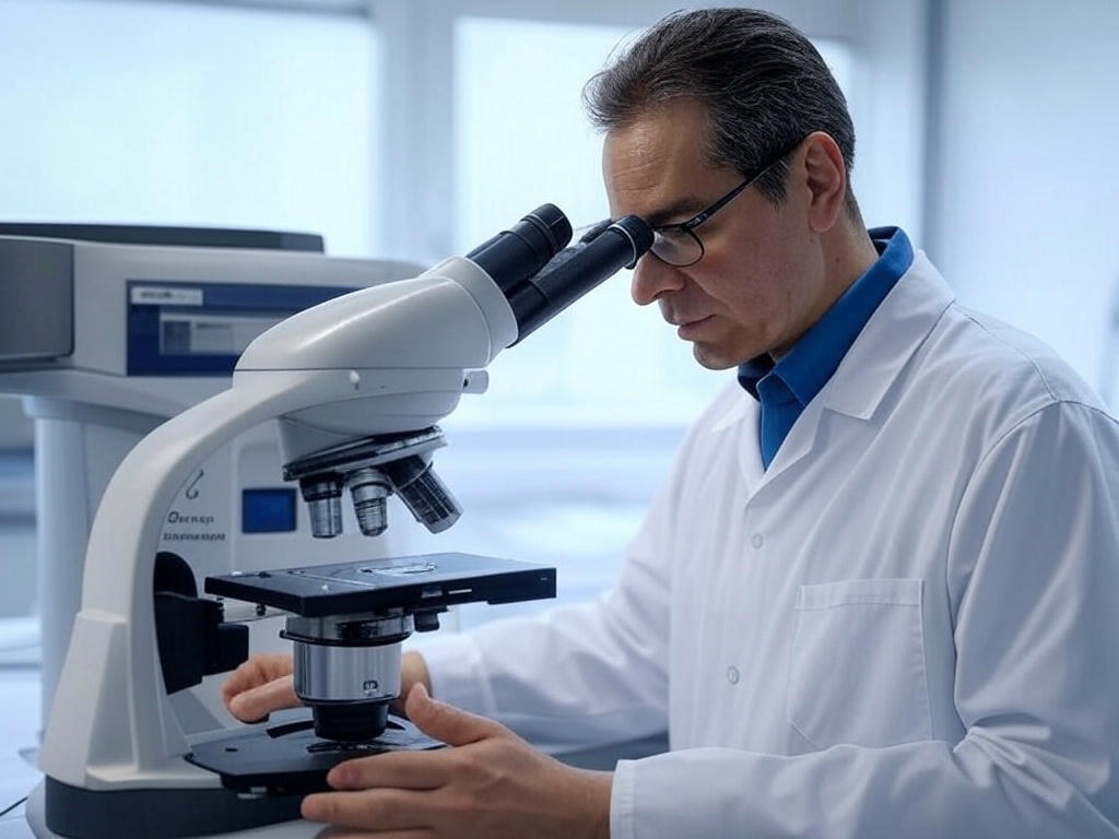
SEM & FESEM Imaging Services
Scanning Electron Microscopy (SEM) and Field Emission SEM (FESEM) are used to capture high-resolution images of a sample's surface and microstructure at very small scales, down to nanometers.
How it Works:
- SEM Sample Preparation: Samples are often coated with a thin layer of conductive material to reduce charging effects during imaging.
- FESEM Imaging: In FESEM, the electron source is a field emission gun (FEG) which produces a more focused beam of electrons compared to traditional SEM, leading to higher resolution imaging.
- EDX (Energy Dispersive X-ray) Analysis: This provides elemental composition data of the sample surface:
- Point Analysis: Measures the elemental composition at a specific point on the sample.
- Line Scan: Provides a line profile of elemental distribution across a specific path.
- Mapping: Creates a map of elemental distribution over a defined region of the sample surface.
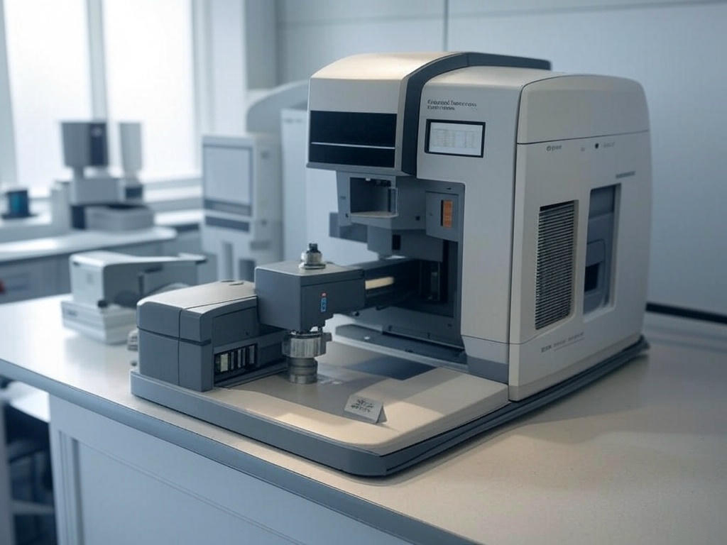
- Floor-type SEM Imaging: This setup is used for larger samples, allowing easier access to large or heavy materials that cannot fit in standard SEM chambers.
- Tabletop SEM Imaging: Compact and affordable, this SEM variant is designed for smaller samples, providing ease of use and portability with optional EDS analysis for material characterization.
Expected Outcomes:
- High-resolution imaging of the sample's surface structure.
- Elemental composition of materials with pinpoint accuracy.
- Ability to identify surface features such as fractures, coatings, and defects.
Visualize your materials at a microscopic level. Contact us for SEM & FESEM Services.

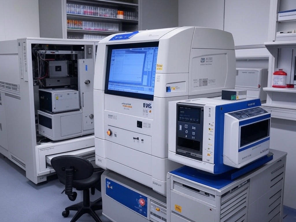
X-ray Imaging Services
X-ray imaging allows you to view both the external and internal structures of your sample without any need for destructive preparation. We offer both 2D and 3D X-ray imaging for comprehensive analysis.
How it Works:
- 2D X-ray: This technique produces traditional radiographic images, capturing a flat, single-view image of the object or sample.
- 3D X-ray (CT or Computed Tomography): This advanced method takes multiple X-ray images from different angles and uses computer processing to reconstruct a 3D model of the sample. It’s ideal for understanding internal structures like voids, cracks, and assembly details.
Expected Outcomes:
- Non-destructive analysis of both internal and external features.
- Precise 3D reconstructions to visualize internal features like defects, voids, and inclusions.
- Ideal for inspecting the quality of components without damaging the sample.
Investigate internal structures and defects with no sample damage. Schedule your X-ray Imaging.
AFM & Optical Microscopy
Atomic Force Microscopy (AFM) and Optical Microscopy are powerful tools for imaging and analyzing the topography and mechanical properties of materials at micro and nano scales.
How it Works:
- AFM: AFM uses a sharp tip that scans the surface of the material to measure its topography at an atomic level. It can also measure other properties like stiffness, adhesion, and friction.
- Optical Microscopy: Uses visible light and lenses to magnify the surface features of the sample, suitable for imaging samples that are transparent or semi-transparent, and for general material inspections.
Expected Outcomes:
- High-resolution surface images and topography at the nano-scale.
- Quantitative data on mechanical properties like hardness, roughness, and elasticity.
- Clear optical images for identifying macro-scale surface features such as cracks or uniformity.
Get insights into your material's properties at various scales. Request AFM or Microscopy Analysis.
AES & TOFSIMS Surface Analysis
Auger Electron Spectroscopy (AES) and Time-of-Flight Secondary Ion Mass Spectrometry (TOF-SIMS) are techniques used to analyze the chemical composition and depth profiling of the surface of materials.
How it Works:
- AES: This technique uses electron bombardment to excite surface atoms, causing them to emit Auger electrons, which are then detected to determine the elemental composition of the surface.
- TOF-SIMS: This technique uses a focused ion beam to dislodge secondary ions from the sample surface, which are then analyzed to provide chemical information about the sample’s surface and its depth profile.
Expected Outcomes:
- Detailed surface chemical composition, including elemental and molecular species.
- Depth profiling to analyze changes in chemical composition below the surface.
- Ability to detect and quantify thin films, surface modifications, and contamination layers.
Gain detailed surface chemical insights and composition data. Contact us for AES & TOFSIMS.
Frequently Asked Questions (FAQ)
What is the difference between SEM and FESEM?
SEM uses a traditional electron gun, while FESEM uses a field emission gun (FEG), allowing for better resolution and imaging of smaller features due to its more focused electron beam.
How long does an imaging session typically take?
The time required for imaging depends on the complexity of the sample and the technique used. SEM and FESEM imaging can take from 1 to several hours, while X-ray imaging may take longer depending on the 3D reconstruction process.
Are there any special sample preparation requirements?
For SEM and FESEM, samples often need to be coated with a conductive layer to prevent charging effects. Other techniques, such as X-ray and AFM, may have different requirements depending on the sample's nature.
Can you perform 3D analysis with X-ray imaging?
Yes, 3D X-ray imaging (CT) is specifically designed to provide a detailed three-dimensional reconstruction of a sample, allowing you to view internal features without damaging the sample.
What types of materials can you analyze?
We can analyze a wide variety of materials including metals, plastics, ceramics, composites, biological tissues, and more. Each technique is tailored to suit the material being examined.
Do you offer post-analysis consultation?
Yes, our experts provide detailed analysis reports and can offer consultation services to help interpret the data obtained from our imaging techniques.